Physiology in sleep
The behavioural inactivity which characterises sleep hides a variety of
physiological events that researchers have been able to reveal, thanks to
the recording of the bioelectric activity of the brain
(electroencephalogram![]() ),
dskeletal muscles (electromyogram
),
dskeletal muscles (electromyogram![]() ),
the hearth (electrocardiogram
),
the hearth (electrocardiogram![]() )
and the mechanical activity of the respiratory muscles (Respyrogram/Spyrogram
)
and the mechanical activity of the respiratory muscles (Respyrogram/Spyrogram![]() ).
).
The recording of all these variables together is know as
polysomnography![]() .
.
The table below shows the changes in activity
(compared to the waking state) of the
vegetative
nervous system![]() in its two parts:
sympathetic
in its two parts:
sympathetic![]() (which predisposes the organism to action) and
parasympathetic
(which predisposes the organism to action) and
parasympathetic![]() (which predisposes the organism to rest) and the cardiocirculatory,
respiratory and thermoregulatory functions observed in
NREM
(which predisposes the organism to rest) and the cardiocirculatory,
respiratory and thermoregulatory functions observed in
NREM![]() and
REM
and
REM![]() stages of sleep.
stages of sleep.
|
Function |
NREM Sleep |
Sonno REM |
| vegetative | prevalence of parasympathetic activity | irregular sympathetic and parasympathetic activity |
| cardiocircolatory | reduced | irregular |
| respiratory | reduced | irregular |
| thermoregulatory | normal | depressed or suppressed |
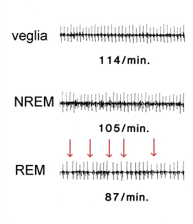 Fig. 1: Electrocardiogram recorded during the stages of the sleeping-waking cycle of animals. The vertical sections show the heart beats. Under each line we can see the cardio frequency (beats/min). As indicated by the arrows, during REM sleep the heart beat pattern is very irregular (Credit: A. Azzaroni e P.L. Parmeggiani.) |
In NREM sleep, cardiocirculatory activity drops compared to the waking state and cardio frequency is reduced (Fig. 1). This is due to an increase in the activity of the parasympathetic section and a decrease in the activity of the sympathetic section of the vegetative nervous system. This is perfectly in line with the condition of reduced motor activity that characterises NREM sleep. In contrast, in REM sleep the cardiocirculatory activity is quite variable, due to the effect of marked irregularities in the vegetative nervous system. As indicated by the arrows in Fig. 1, the heart beat is much less regular.
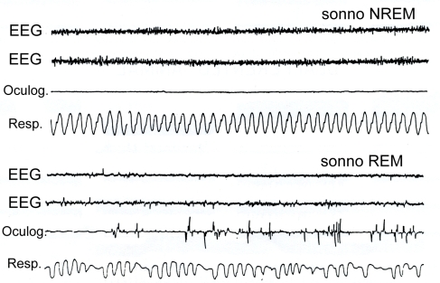 Fig. 2:
: Polysomnography effected during NREM sleep and REM sleep. |
In NREM sleep, breathing is regular (Fig. 2). The breathing rate is
less frequent compared to the waking state because the resting muscles
reduce the organism’s need for oxygen. In REM sleep, instead, the
frequency and breadth of ventilation are irregular, especially in
association with rapid eye movements and
mioclonie
|
|
|
Thanks to thermoregulation, "warm blooded" animals (omeotermi![]() )
manage to keep their body temperature unchanged even in unfavourable
weather conditions. At low temperatures, their body temperature is
kept constant by cutaneous
vasoconstriction
)
manage to keep their body temperature unchanged even in unfavourable
weather conditions. At low temperatures, their body temperature is
kept constant by cutaneous
vasoconstriction![]() and the presence of muscular
joilts
and the presence of muscular
joilts![]() ,
and at high temperatures by cutaneous
vasodilatation
,
and at high temperatures by cutaneous
vasodilatation![]() and, in addition, sweating in man or
thermic polypnea
and, in addition, sweating in man or
thermic polypnea
![]() in animals.
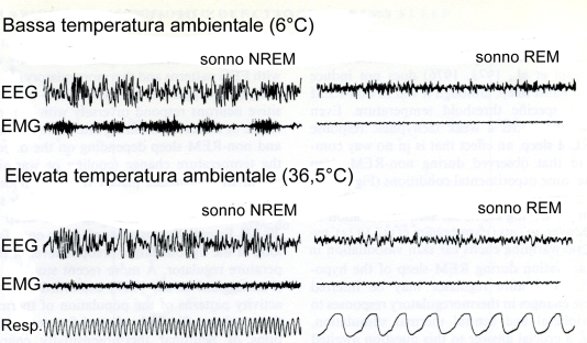
These thermoregulatory responses are present in NREM sleep, while they
are depressed or suppressed in REM sleep. As we can see in Fig. 3, in an
animal kept at low temperatures, the jolt recorded by the EMG during NREM
sleep (A) disappears during REM sleep (B). In the same way, in the animal
kept at high temperatures, thermic polypnea, indicated by a high frequency
breathing rate during NREM, disappears during REM sleep. Although some of
the mechanisms that lead to the suspension of thermoregulation during REM
sleep have been understood, we still do not really know why this happens.
in animals.
These thermoregulatory responses are present in NREM sleep, while they
are depressed or suppressed in REM sleep. As we can see in Fig. 3, in an
animal kept at low temperatures, the jolt recorded by the EMG during NREM
sleep (A) disappears during REM sleep (B). In the same way, in the animal
kept at high temperatures, thermic polypnea, indicated by a high frequency
breathing rate during NREM, disappears during REM sleep. Although some of
the mechanisms that lead to the suspension of thermoregulation during REM
sleep have been understood, we still do not really know why this happens.
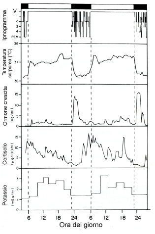 |
Endocrine secretions also undergo complex changes, depending on the different stages of sleep. For example, as we can see in Fig. 4, where the waking period is shown by the white bar and the sleeping period by the black bar, the hypophysial growth hormone is secreted mostly in the initial stages of sleep during the NREM part, while the secretion of cortisolo increases during the end of the sleeping period and on awaking.
Fig. 4: Changes in human body temperature over two consecutive days,
and concentration in the plasma of the growth hormone, cortisolo and
potasium. At the top, the waking period is shown by the white bar and
the sleeping period by the black bar and by the line. During this period
we can also see how the stages of sleep alternate
(hypnogram |