The Brain
The human brain weighs on
average 1500
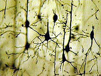
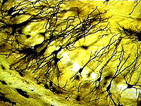
grams. It is estimated that it contains approximately 100 billion primary cells (neurons![]() ) and most likely a similar number of supporting cells (glial
cells). A typical neuron is composed of a cell body and several expansions, of
which the shorter ones (dendrites
) and most likely a similar number of supporting cells (glial
cells). A typical neuron is composed of a cell body and several expansions, of
which the shorter ones (dendrites![]() ) receive connections and signals from other
neurons, while the longest one (axon
) receive connections and signals from other
neurons, while the longest one (axon![]() ) forms in turn connections with other
neurons.
) forms in turn connections with other
neurons.
|
Fig. 1:
Hippocampal |
 |
 |
A typical neuron receives from
several hundreds to tens of thousands of connections (synapses![]() ) and forms in turn
a variable number of them.
) and forms in turn
a variable number of them.
|
Fig. 2: Nerve impulse for neurotransmission. (Credit: Eric H. Chudler - "Neuroscience for kids") These connections are the essential structures for the formation of complex nerve circuits along which information travels. Primarily, the neural information system uses electrical signals (the fastest of which is the action potential, Fig. 2), which are not usually able to jump from neuron to neuron, because there is no physical continuity among cells at the level of their connections, called synapses. |
|
 Fig. 4: Release of neurotransmitter from vesicles containing it (red dots) at the pre-synaptic terminal. (Credit: Eric H. Chudler - "Neuroscience for kids") |
At the synapse, therefore, the electrical signal promotes the release of a
chemical signal (in the form of molecules called neurotransmitters![]() )
that elicits again an electrical signal in the next neuron (Fig. 4).
)
that elicits again an electrical signal in the next neuron (Fig. 4).
Today, we know in depth
about the main cellular and
molecular events of neural activity, but we have a much less detailed
knowledge of how neural circuits work when they are performing complex cerebral
functions. However, non-invasive imaging techniques able to monitor brain
activity (Magnetic
Nuclear Resonance![]() ,
Positron Emission Tomography
,
Positron Emission Tomography![]() ,
and others) are evolving very rapidly. These techniques will allow us, in a near
future, to study neuropathological states much better and also to understand
much better how brain circuits work. Fig. 5 is an example of how, by using these
methods, it is possible to monitor which areas of the cerebral cortex are active
(red color) when a subject is looking a figure.
,
and others) are evolving very rapidly. These techniques will allow us, in a near
future, to study neuropathological states much better and also to understand
much better how brain circuits work. Fig. 5 is an example of how, by using these
methods, it is possible to monitor which areas of the cerebral cortex are active
(red color) when a subject is looking a figure.
 |
Fig. 5: Magnetic Resonance
Imaging (MRI) from a human brain. The primary visual cortex is active while
the subject is looking at a figure. (Credit: J.Cohen, University of Pittsburgh and Carnegie Mellon University e N.Goddard, Pittsburgh Supercomputing Center - "Watching the brain in action") |